
Characterizing molecular recognition and regulation events involving signalosomes in the retina
#retinasignalosomes
Description
One of the outstanding mysteries of visual perception lies in the precise functions of the intricate molecular circuits of the retina. Synaptic contacts between different cell types enable transformation of light signals into fine chemical messages, ultimately reaching our brain. However, for many of these key macromolecules, our current knowledge is extremely limited. In our project we use the most advanced in vitro technologies to characterize the structures, interactions and functions of recently discovered protein networks called ” signalosomes”, essential for visual perception, particularly for night vision. Our aim is to collect insights to provide an accurate molecular mapping of the interactions underlying these fundamental proteins and, at the same time, to elucidate the molecular defects causing rare untreatable vision diseases such as congenital stationary night blindness. We expect that our results will pave the way to exciting new opportunities for usage and harnessing of these molecules in future bio-medical applications.
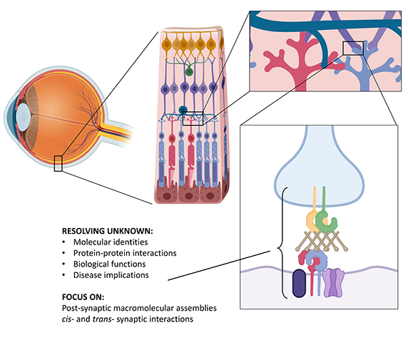 |
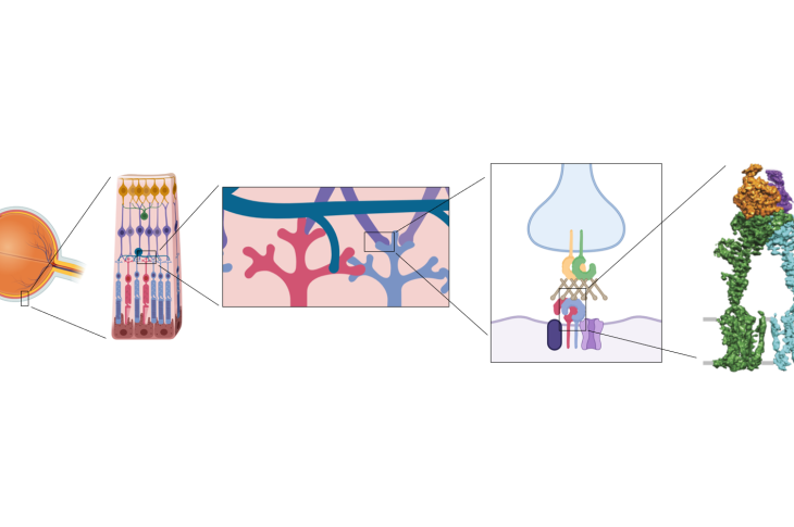
The figure shows a summary scheme of our investigation targets in the retina, at the synaptic interface between photoreceptors and bipolar cells. |
Pikachurin and its interactions with GPR179 – In the retina, photoreceptors form synaptic contacts with ON-type bipolar cells to ensure efficient synaptic transmission. The extracellular matrix proteoglycan Pikachurin, a distroglycan ligand with features strongly resembling to those of its widespread homolog agrin, connects a presynaptic complex on the photoreceptor with the postsynaptic orphan receptor GPR179. We tackled the structural and functional characterization of Pikachurin N- and C-terminal fragments, and in collaboration with the group of Kirill Martemyanov at Scripps Florida we studied the extracellular molecular contacts enabling the interaction between Pikachurin C-terminal EGF3 domain and the ectodomain of GPR179. Our findings provide insights into how pikachurin precisely positions GPR179 at synapses to ensure neurotransmission.
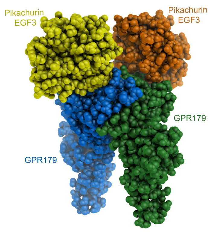 |
The figure shows a cartoon representation of the CryoEM reconstruction of the extracellular fragments of dimeric GPR179 (blue and green) forming 2:2 complexes with the C-terminal EGF3 domains of human Pikachurin. |
Resources
PDB Files:
- 7ZCB – Crystal structure of Human Pikachurin/EGFLAM N-terminal Fibronectin-III (1-2) domains
- 7ZC9 – Crystal structure of Human Pikachurin/EGFLAM C-terminal Laminin-G domain (LG3)
- 8D1B – CryoEM structure of human orphan GPCR GPR179 in complex with extracellular matrix protein pikachurin
EMDB Files:
- EMD-27121 – CryoEM structure of human orphan GPCR GPR179 in complex with extracellular matrix protein pikachurin
SASBDB Files:
Publications and Dissemination
Publications
- Patil D.N.*, Pantalone S., Cao Y., Laboute T., Novick S.J., Singh S., Savino S., Faravelli S., Magnani F., Griffin P.R., Singh A.K., Forneris F.*, Martemyanov K.A.* (*Shared Corresponding Authors). Structure of the photoreceptor synaptic assembly of the extracellular matrix protein pikachurin with the orphan receptor GPR179. Science Signaling, 16, eadd9539 (2023). – PubMed – UNIFIND
Press releases
- Joint University of Pavia press release about Patil et al., Science Signaling (2023): PDF in Italian