
Other protein studies from our lab
This section provides a summary of targets studied that do not fall within the main lab's research topics
Description
Receptor tyrosine kinase-like orphan receptor 1 (hROR1)
Receptor tyrosine kinase-like orphan receptors (RORs) are poorly characterized receptor tyrosine kinases involved in embryonic development and muscle differentiation. Their importance is highlighted by ROR gene overexpression during cancer progression, particularly in solid and hematopoietic malignancies. Because ROR1 expression is strongly restricted in cancer cells and remains low in healthy adult tissues, it represents an attractive marker for targeted cancer therapy. Alongside drug-discovery campaigns targeting the intracellular kinase domains, antibodies directed against the kringle domains of human ROR1 and ROR2 have shown selective antitumor activity. To clarify the extracellular architecture of these receptors, we produced, crystallized, and determined the high-resolution structure of the isolated kringle domain of human ROR1 (hROR1-KRD).
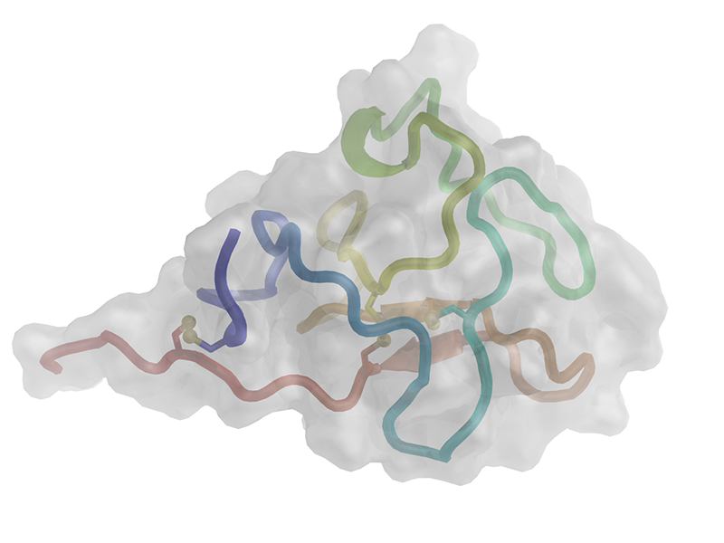 |
Cartoon representation of hROR1-KRD. The polypeptide chain is colored from the N-terminus (blue) to the C-terminus (red). Disulfide bonds are shown as balls and sticks, and residues defining the boundaries of the secondary-structure elements are labeled. |
Resources
PDB Files:
- 7TNG – Kringle domain of human receptor tyrosine kinase-like orphan receptor 1 (ROR1)
Companion files are available in our data repository.
Publications and Dissemination
Publications
- Guarino S.R., Di Bello A., Palamini M., Capillo M.C., Forneris F. Crystal structure of the kringle domain of human receptor tyrosine kinase-like orphan receptor 1 (hROR1). Acta Crystallographica section F – Structural Biology Communications, F78, 185-192 (2022). – PubMed – UNIFIND
Clinical-grade immunocytokines
Through a collaboration with Dr Alessandra Villa (Philochem AG) and Prof. Dario Neri (ETH Zurich), we contributed to the structural characterization of clinical-grade immunocytokines. We determined the three-dimensional structure of the common L19 diabody fragment and used SEC-SAXS and negative-stain electron microscopy to characterize three flexible full-length immunocytokines. These results show that low-resolution structural characterization provides valuable complementary information for immunocytokine quality control and can guide the design and optimization of these chimeric protein reagents for clinical development.
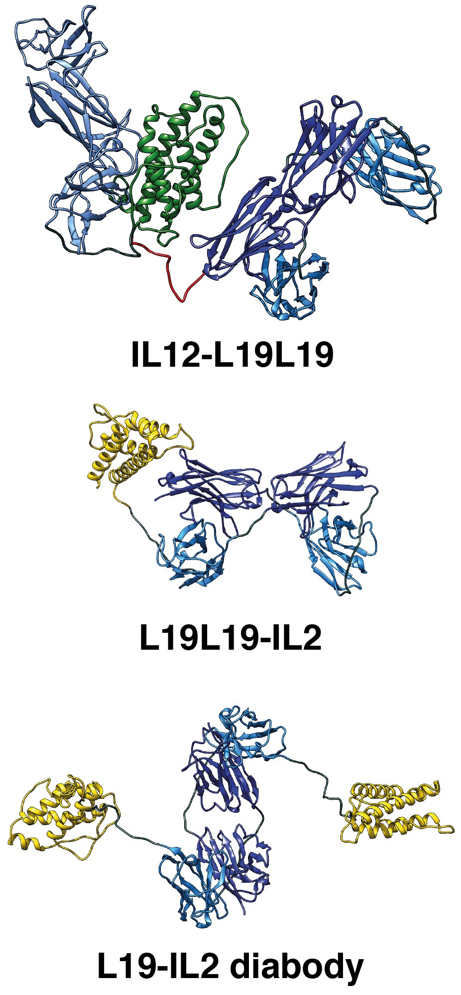 |
Cartoon representation of the molecular models of L19-IL2, L19L19-IL2, and IL12-L19L19 immunocytokines derived from SEC-SAXS data. |
Resources
PDB Files:
- 7AH1 – L19 diabody fragment from the L19-IL2 immunocytokine
SASBDB Files:
- SASDJN8 – L19-IL2 dimeric immunocytokine measured using SEC-SAXS
- SASDJP8 – L19L19-IL2 monomeric immunocytokine measured using SEC-SAXS
- SASDJQ8 – IL12-L19L19 monomeric immunocytokine measured using SEC-SAXS
Companion files are available in our data repository.
Publications and Dissemination
Publications
- Ongaro T.*, Guarino S.R.*, Scietti L., Palamini M., Wulhfard S., Neri D., Villa A.**, Forneris F.** (*Equal Contribution – **Shared Corresponding Authors) Inference of molecular structure for characterization and improvement of clinical grade immunocytokines. Journal of Structural Biology, 213, 107696 (2021). – PubMed – UNIFIND
LOG phosphoribonucleotide hydrolases
Through a collaboration with Prof. Ivo Frébort and Prof. Petr Galuszka (Palacký University Olomouc, Czech Republic), we contributed to the structural characterization of a recently discovered protein class named “Lonely Guy” (LOG). These enzymes are central to cytokinin biosynthesis because they convert inactive cytokinin nucleotides into biologically active free bases. We determined the three-dimensional structure of a fungal LOG protein from Claviceps purpurea in ligand-free and ligand-bound states. Our structural and biochemical analyses clarified substrate recognition and identified an accurate sequence signature for distinguishing Rossmann-fold LOG-like phosphoribonucleotide-processing enzymes from misannotated lysine decarboxylases.
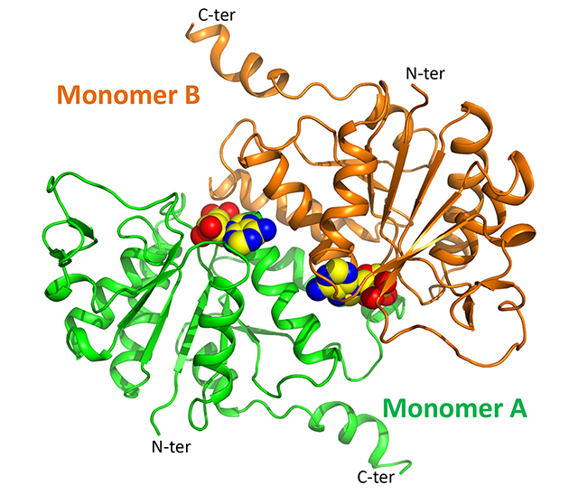 |
Overview of the three-dimensional structure of cpLOG, rendered as a cartoon. The reaction product phosphoribose, identified in the catalytic pocket, is shown as spheres. |
Resources
PDB Files:
- 5AJT – Ligand-free phosphoribohydrolase Lonely Guy from Claviceps purpurea
- 5AJU – Phosphoribohydrolase Lonely Guy from Claviceps purpurea in complex with phosphoribose
Companion files are available in our data repository.
Publications and Dissemination
Publications
- Dzurová L., Forneris F.*, Savino S., Galuszka P., Vrabka J., Frébort I.* (*Shared Corresponding Authors) The three-dimensional Structure of Lonely Guy from Claviceps purpurea provides insights into the phosphoribohydrolase function of Rossmann fold-containing lysine decarboxylase-like proteins. Proteins, 83, 1539-1546 (2015). – PubMed – UNIFIND
Targeting Burkholderia cenocepacia quorum-sensing virulence to fight cystic fibrosis
Burkholderia cenocepacia is a rare opportunistic pathogen and a major concern among respiratory infections in people with cystic fibrosis. It is particularly difficult to treat because of its high resistance to clinically relevant antimicrobial agents. Its quorum-sensing cell–cell communication system controls several processes essential for bacterial virulence, including biofilm formation and protease and siderophore production. Targeting the enzymes involved in this pathway therefore represents a promising therapeutic strategy. This research was performed in collaboration with the Molecular Microbiology Group of our Department, led by Prof. Giovanna Riccardi.
DfsA. The bifunctional crotonase DfsA is a key component of the B. cenocepacia quorum-sensing pathway. We determined the three-dimensional structure of the enzyme in complex with the non-physiological reaction product lauric acid. Biochemical characterization showed that, unlike the physiological product BDSF (Burkholderia diffusible signaling factor, cis-2-dodecenoic acid), lauric acid is released slowly and behaves as an inhibitory end product. The structure explains how DfsA accommodates long aliphatic substrate chains without requiring the previously proposed conformational changes and provides a template for structure-based development of antivirulence compounds.
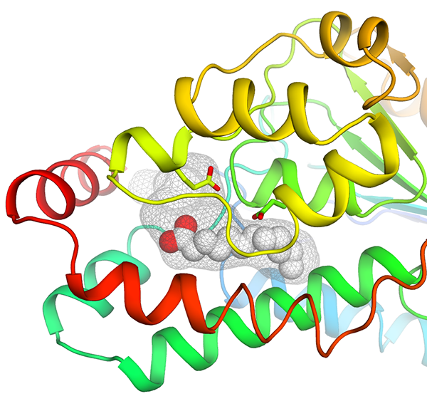 |
Cartoon representation of the DfsA structure, colored from the N-terminus (blue) to the C-terminus (red). The deep hydrophobic cavity is shown as a grey mesh, and the non-physiological reaction product lauric acid is shown as spheres near the catalytic pocket. |
CepI. Our collaborators identified effective non-competitive inhibitors of the B. cenocepacia acyl-homoserine-lactone synthase CepI. Because CepI resisted crystallization both without and with these inhibitors, we combined homology modelling and in silico docking. Site-directed mutagenesis and biochemical assays then validated the predicted binding sites and clarified the mechanism of action of these candidate drugs.
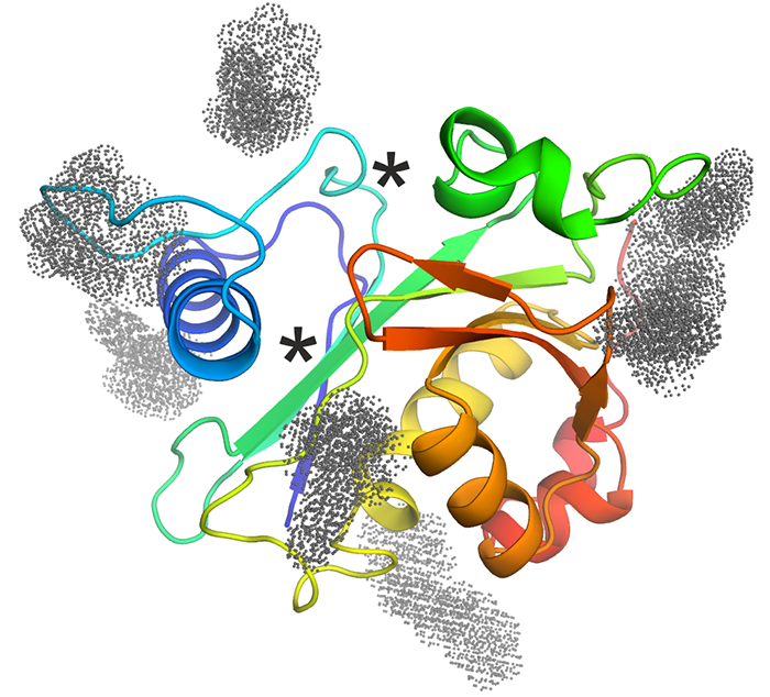 |
Cartoon representation of the CepI homology model, colored from the N-terminus (blue) to the C-terminus (red). Known substrate-binding sites are marked with asterisks, and optimized inhibitor-binding sites identified by molecular docking are shown as colored dots. |
Resources
PDB Files:
- 5FUS – Crystal structure of Burkholderia cenocepacia DfsA
Companion files are available in our data repository.
Publications and Dissemination
Publications
- Buroni S.*, Scoffone V.C.*, Fumagalli M., Makarov V., Cagnone M., Trespidi G., De Rossi E., Forneris F., Riccardi G., Chiarelli L.R. (*Equal Contribution) Investigating the Mechanism of Action of Diketopiperazines Inhibitors of the Burkholderia cenocepacia Quorum Sensing Synthase CepI: A Site-Directed Mutagenesis Study. Frontiers in Pharmacology, 9, 836 (2018). – PubMed – UNIFIND
- Spadaro F.*, Scoffone V.C.*, Chiarelli L.R.**, Fumagalli M., Buroni S., Riccardi G., Forneris F.** (*Equal Contribution – **Shared Corresponding Authors) The crystal structure of Burkholderia cenocepacia DfsA provides insights into substrate recognition and quorum sensing fatty acid biosynthesis. Biochemistry, 55, 3241-3250 (2016). – PubMed – UNIFIND
- Scoffone V.C.*, Chiarelli L.R.*, Makarov V.*, Brackman G., Israyilova A., Azzalin A., Forneris F., Riabova O., Savina S., Coenye T., Riccardi G., Buroni S. (*Equal Contribution) Discovery of new diketopiperazines inhibiting Burkholderia cenocepacia quorum sensing in vitro and in vivo. Scientific Reports, 6, 32487 (2016). – PubMed – UNIFIND
- Israyilova A., Buroni S., Forneris F., Scoffone V.C., Shixaliyev N.Q., Riccardi G., Chiarelli L.R. Biochemical characterization of Glutamate Racemase, a new candidate drug target against Burkholderia cenocepacia infections. PLoS One, 11, e0167350 (2016). – PubMed – UNIFIND